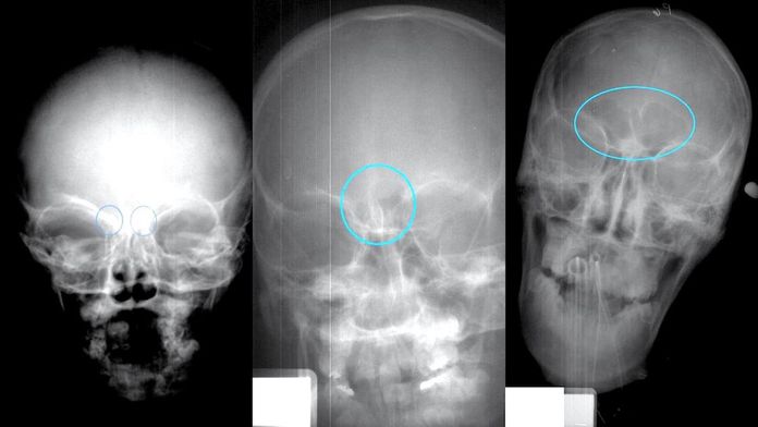
In a what they are calling a “proof-of-concept” study, anthropologists from North Carolina State University have developed a new technique using frontal sinus growth to estimate age at death in juvenile populations.
The sinus cavity is formed during childhood as a portion of the frontal bone, the bone that forms the human forehead, separates over time, kind of like a puff pastry. In science talk, the ectocranial table, the skin side of the bone, separates from the endocranial table, the brain side of the bone, forming an air pocket, the frontal sinus. The endocranial table (brain side) stops growing when the brain stops growing, while the skin side portion of the frontal bone continues to grow as the face grows.
To turn sinus growth into an age estimation technique, Kaitlin Moore, former NCSU graduate student, and Professor Ann Ross examined frontal bone X-rays taken from 392 individuals ranging in age from 0 to 18. Moore and Ross grouped the images based on sinus cavity growth, eventually quantifying four distinct stages of development. The pair tied the developmental stages to age ranges through statistical analyses (one-way ANOVA) and created a regression equation to test how well developmental stage predicts age. The technique proved statistically sound, though Moore and Ross caution that age ranges provided are broad. As Ross stated in a press release,
"The next step is to look at a larger sample of X-rays to further fine-tune the technique. It's useful now, but it would be even more valuable if we could improve its specificity.”
Moore and Ross published their findings in the June edition of the Anatomical Record.
Moore, K. and Ross, A. (2017), Frontal Sinus Development and Juvenile Age Estimation. Anat. Rec.. doi:10.1002/ar.23614


